


Not sure what to say to someone with depression? This in-depth guide breaks down 7 compassionate, practical...
Waterfall is one of the most useful late-game HMs in Pokémon X and Y, but many players...
Want to connect your Pinterest boards with your Facebook audience? This guide explains how to add a...
Chronic emphysema and chronic bronchitis are both forms of COPD, but they affect the lungs in different...
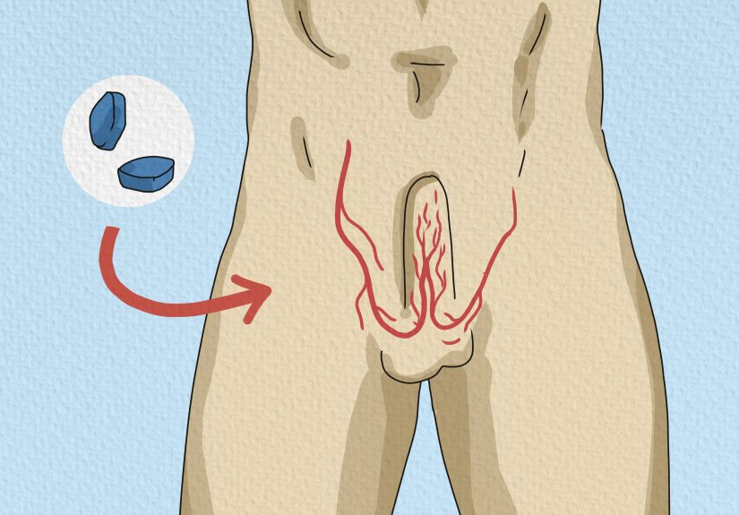
Wondering how to tell if a man is taking Viagra? This guide explains six possible signs, including...

What do workers know that customers usually do not? This fun, practical guide reveals 50 little industry...

Want your house to become the one trick-or-treaters talk about all night? An 18-foot Halloween serpent inflatable...

Spoon nails can look like a minor cosmetic issue, but they often tell a bigger health story....

A pedestrian car accident can turn an ordinary walk into a life-changing emergency in seconds. This in-depth...

Interested in a coworker but want to keep things classy, clear, and professional? This guide breaks down...