Table of Contents >> Show >> Hide
- Why Imaging Matters in Gaucher Disease
- The Radiology Lineup: What Each Test Does Best
- X-Ray (Plain Radiographs): Fast, Cheap, and Surprisingly Useful
- MRI: The MVP for Bone Marrow and Early Complications
- DEXA/DXA (Bone Density Scan): “How Strong Are the Bones?”
- CT: High Detail, Useful for Specific Questions (But Mind the Radiation)
- Ultrasound: The No-Radiation Workhorse for Organ Size
- Nuclear Medicine (Bone Scan, PET): Occasionally Helpful, Not a Daily Driver
- What Radiologists Look for in Gaucher Bones
- MRI in Gaucher Disease: Beyond “Yes/No” Imaging
- CT, MRI, and Ultrasound for Liver and Spleen
- Chest Imaging: When Lungs Get Involved
- A Sensible Imaging Strategy (General Education, Not Personal Medical Advice)
- Scan-Day Tips: How to Make Imaging Less Miserable (and More Useful)
- Real-World Experiences: What People Often Notice About Gaucher Imaging (Extra Depth)
- Key Takeaways
Gaucher disease is a rare genetic (lysosomal storage) disorder where certain fatty substances build up inside cellsespecially in the spleen, liver, and bone marrow.
If you’re living with Gaucher, or caring for someone who is, you’ll quickly learn a truth that’s equal parts annoying and empowering:
you can’t “eyeball” Gaucher. Symptoms can be subtle, labs don’t tell the whole story, and bones can be quietly changing long before they start loudly complaining.
That’s where radiology comes in. Imaging tests like X-ray, MRI, DXA/DEXA, CT, and ultrasound help clinicians:
confirm the extent of organ and skeletal involvement, detect complications early (hello, avascular necrosis),
track treatment response, and make smarter decisions about monitoring over time.
This article breaks down what each scan can (and can’t) show in Gaucher diseaseusing real-world, practical explanations in plain American English,
with just enough humor to keep things readable (because the MRI machine already provides enough drama).
Why Imaging Matters in Gaucher Disease
Gaucher disease affects multiple organ systems, but radiology is especially helpful for two big domains:
(1) the skeleton and (2) visceral organs (mainly the spleen and liver).
Bones are a major focus because Gaucher-related marrow infiltration can trigger a cascade of problems: decreased bone density, infarcts, painful “bone crises,” fractures,
and osteonecrosis (bone tissue damage related to reduced blood supply).
Imaging helps answer questions clinicians can’t reliably answer with symptoms alone:
Is bone pain coming from active marrow disease, a fracture, osteonecrosis, or something unrelated?
Is the spleen simply enlargedor are there nodules that need closer follow-up?
Is treatment stabilizing bone marrow infiltration over time?
The Radiology Lineup: What Each Test Does Best
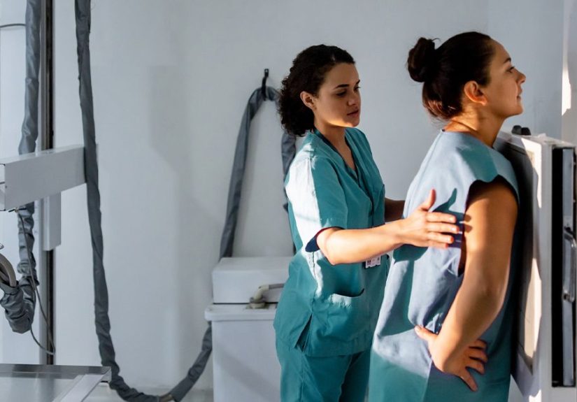
X-Ray (Plain Radiographs): Fast, Cheap, and Surprisingly Useful
X-rays are often the “first look” when someone has bone pain, suspected fracture, joint collapse, or a new deformity.
They’re quick and accessible, but they don’t show bone marrow infiltration wellso X-ray is great for the “hard structure” story,
not the “what’s happening inside the bone” story.
- Best for: fractures, bone deformities, joint space changes, advanced osteonecrosis/collapse, certain classic Gaucher bone shapes
- Limitations: can miss early osteonecrosis and marrow disease; normal X-ray does not rule out significant Gaucher bone involvement
A classic radiographic pattern associated with Gaucher is the Erlenmeyer flask deformitya flaring of the end of long bones
(often the distal femur) due to abnormal bone remodeling. It’s not exclusive to Gaucher, but it’s a recognizable clue, especially in the right clinical context.
MRI: The MVP for Bone Marrow and Early Complications
If Gaucher radiology had a “most valuable player,” MRI would be wearing the crown (and probably earplugs, because MRI scanners are loud).
MRI excels at evaluating bone marrow infiltration, detecting early osteonecrosis, and assessing
the extent of skeletal involvementoften before X-rays show much at all.
- Best for: marrow infiltration, osteonecrosis (early detection), infarcts, marrow edema, spine/femur monitoring, whole-body assessment
- Limitations: longer exam time, motion sensitivity, can be difficult with claustrophobia; sedation may be needed for some children
- Radiation: none (MRI does not use ionizing radiation)
MRI findings in Gaucher often reflect marrow replacement/infiltration patterns.
Radiologists may use standardized scoring systems to quantify severity and monitor changes over time (more on that soon).
DEXA/DXA (Bone Density Scan): “How Strong Are the Bones?”
DEXA (also written DXA) measures bone mineral density (BMD), commonly at the lumbar spine and hips.
It’s quick, painless, and uses a very low dose of radiationso it’s widely used to evaluate osteopenia and osteoporosis,
both of which can occur in Gaucher due to marrow involvement and altered bone remodeling.
- Best for: tracking bone density (BMD), estimating fracture risk, monitoring osteopenia/osteoporosis over time
- Limitations: does not show marrow infiltration, infarcts, or osteonecrosis; it’s a “density meter,” not a full skeletal disease map
- Pro tip: in younger patients, clinicians often look closely at Z-scores (age-matched), not just T-scores
CT: High Detail, Useful for Specific Questions (But Mind the Radiation)
CT provides detailed cross-sectional images and can be valuable for evaluating organs and complications,
including some lung findings and abdominal anatomy. It can also measure liver and spleen volumes,
but CT uses more radiation than X-ray or DEXAso clinicians typically reserve it for situations where CT offers a clear advantage
or MRI isn’t feasible.
- Best for: abdominal anatomy, splenic/liver assessment when MRI isn’t available, lung evaluation (high-resolution CT), certain complications
- Limitations: radiation exposure; not as sensitive as MRI for marrow infiltration
Ultrasound: The No-Radiation Workhorse for Organ Size
Ultrasound is often used to assess liver and spleen size, especially in children or when frequent monitoring is needed.
While ultrasound is operator-dependent and less comprehensive than MRI for some questions,
it’s a practical and radiation-free option, and some centers use advanced approaches (including 3D ultrasound)
for tracking organ volumes.
- Best for: screening and monitoring hepatosplenomegaly, follow-up checks, pediatric assessments
- Limitations: less detail than MRI for marrow disease; may not fully characterize complex splenic lesions
Nuclear Medicine (Bone Scan, PET): Occasionally Helpful, Not a Daily Driver
Bone scans can detect areas of increased bone turnover and may help in certain diagnostic puzzles (like unclear pain or suspected multifocal lesions),
but they’re not specific for Gaucher. PET is not routine for Gaucher monitoring, but may appear in workups when clinicians are evaluating complications
or other conditions.
What Radiologists Look for in Gaucher Bones
1) Bone Marrow Infiltration
Gaucher cells can accumulate in bone marrow, altering normal marrow composition and function.
MRI is the best tool to visualize the distribution and severity of marrow involvement, commonly focusing on the spine, pelvis, and femurs.
These patterns can correlate with symptom burden and help guide monitoring decisions.
2) Erlenmeyer Flask Deformity
On X-ray, the distal femur (and sometimes other long bones) can appear flared with reduced normal taperingan “Erlenmeyer flask” shape.
It reflects abnormal modeling during growth and may be seen in patients with Gaucher, particularly if skeletal involvement began early in life.
It’s a clue, not a standalone diagnosis.
3) Bone Infarcts and “Bone Crises”
A “bone crisis” in Gaucher can involve severe pain and inflammation, sometimes related to infarction (reduced blood supply) within bone.
MRI may show marrow edema and changes consistent with infarct patterns, helping distinguish an acute skeletal event from other causes of pain.
This matters, because management and urgency can differ depending on what’s actually happening inside the bone.
4) Osteonecrosis (Avascular Necrosis)
Osteonecrosisoften affecting hips and shoulderscan be one of the most serious skeletal complications.
MRI can detect osteonecrosis earlier than X-ray, sometimes before structural collapse occurs.
Early detection can change clinical decisions, including activity recommendations, orthopedic referral timing, and treatment planning.
5) Osteopenia, Osteoporosis, and Fractures
Lower bone mineral density can increase fracture risk.
DEXA provides a standardized way to track bone density over time, while X-ray or CT may identify fractures.
MRI may be useful if pain suggests a fracture that isn’t clearly visible on X-ray, or if clinicians suspect marrow-driven pathology.
MRI in Gaucher Disease: Beyond “Yes/No” Imaging
Whole-Body MRI and Regional MRI
Some centers use targeted MRI (spine + femurs is a common strategy), while others may use whole-body MRI protocols
to map skeletal involvement more broadly.
MRI can also evaluate organs (liver, spleen) when abdominal assessment is neededwithout the ionizing radiation associated with CT.
Scoring Systems: The Bone Marrow Burden (BMB) Score
Gaucher imaging isn’t only about spotting abnormalitiesit’s also about tracking change.
MRI-based scoring systems help quantify skeletal disease and monitor response to therapy.
One widely referenced approach is the Bone Marrow Burden (BMB) score, which provides a semi-quantitative estimate
of marrow involvement, commonly focusing on areas like the lumbar spine and femurs.
Here’s why that’s useful: if a patient starts therapy and symptoms improve, clinicians still want objective evidence that marrow disease is stabilizing.
Conversely, if symptoms worsen, imaging scores can help confirm whether skeletal disease is progressing or whether another issue is driving symptoms.
Example: What Monitoring Might Look Like
Imagine a patient with Gaucher type 1 who has intermittent hip pain and elevated disease burden at baseline.
A baseline MRI shows significant marrow infiltration in the femurs and early changes suspicious for osteonecrosis.
Treatment begins. After a period of therapy and clinical follow-up, a repeat MRI shows improved marrow signal patterns and stable joint architecture.
That imaging stability can be reassuringand it can guide how aggressively clinicians monitor going forward.
CT, MRI, and Ultrasound for Liver and Spleen
Gaucher commonly causes hepatosplenomegaly (enlarged liver and spleen).
Imaging can measure organ size, assess internal structure, and help identify complications such as focal splenic lesions or nodules.
Volumetric measurements (especially with MRI, CT, or advanced ultrasound methods) can be used to track organ response to treatment over time.
- Ultrasound: great for routine size checks and follow-up, especially in pediatrics
- MRI: detailed organ assessment without ionizing radiation; can support volumetric tracking
- CT: detailed anatomy and fast acquisition; useful in specific clinical scenarios but uses higher radiation
A practical point: if imaging detects splenic nodules or unusual lesions, clinicians may recommend follow-up imaging
(often with MRI for better tissue characterization) depending on the patient’s overall clinical situation.
Chest Imaging: When Lungs Get Involved
Lung involvement is less common than bone or organ involvement in many Gaucher cases, but it can occur.
When clinicians suspect pulmonary diseasebased on symptoms like shortness of breath, oxygen issues, or abnormal pulmonary testing
imaging may include chest X-ray and, more importantly, high-resolution CT (HRCT) to evaluate interstitial patterns.
CT can show findings such as interlobular septal thickening and ground-glass changes in reported cases of pulmonary involvement.
A Sensible Imaging Strategy (General Education, Not Personal Medical Advice)
There’s no single imaging schedule that fits everyone with Gaucher disease.
The “right” approach depends on age, symptoms, disease type, treatment status, and prior complications.
That said, published recommendations and expert consensus commonly emphasize:
baseline assessment of skeletal disease (often with MRI and radiographs as indicated),
plus periodic monitoring with MRI and DXA/DEXA in patients with skeletal involvement or risk factors.
Baseline Imaging Often Includes
- MRI (spine and femurs/pelvis): to evaluate marrow infiltration and silent complications
- DEXA/DXA: to establish bone mineral density baseline
- X-rays (targeted): if pain, deformity, suspected fracture, or joint issues are present
- Ultrasound or MRI/CT abdomen: to document liver/spleen size and structure when needed
Follow-Up Imaging Tends to Be Symptom-Driven + Risk-Adjusted
If someone has new focal bone pain, MRI may be prioritized to evaluate osteonecrosis or infarct.
If bone density is low, clinicians may repeat DEXA on an interval appropriate for age and risk.
If organs are markedly enlarged or changing, ultrasound or volumetric MRI/CT may be used for monitoring.
Scan-Day Tips: How to Make Imaging Less Miserable (and More Useful)
For MRI
- Ask about “wide-bore” scanners if claustrophobia is an issue.
- Bring prior imaging reports (or make sure the facility can access them) so radiologists can compare changes over time.
- Know the question: “Rule out osteonecrosis?” “Assess marrow burden?” The exam protocol may differ based on the goal.
For DEXA
- Keep it consistent: Using the same facility/device when possible can make trend comparisons easier.
- Ask which score matters: younger patients often focus on Z-scores rather than T-scores.
For CT and X-ray
- Share your imaging history so clinicians can avoid unnecessary repeat radiation-based studies when alternatives exist.
- Contrast questions: if IV contrast is planned, you may be asked about kidney function and allergies.
Real-World Experiences: What People Often Notice About Gaucher Imaging (Extra Depth)
People dealing with Gaucher disease often describe imaging as a strange mix of reassurance and frustrationreassuring because it provides concrete answers,
frustrating because it’s time-consuming, expensive, and sometimes feels like you’re collecting “receipts” for symptoms you already know are real.
One common experience is learning that pain and imaging don’t always match.
Someone might feel intense bone pain, yet X-rays look normaluntil an MRI reveals marrow edema or early osteonecrosis that plain films simply can’t show.
That moment can feel validating: “Oh, so I’m not imagining this.” It can also be unsettling: “Wait… this has been happening under the surface?”
MRI, in particular, tends to become the main character in the Gaucher story.
Many patients say the first MRI was intimidatingnot because of needles or surgery, but because it’s a commitment:
you lie still in a loud machine that sounds like a robot learning to play drums.
People who do best often come prepared: they ask for headphones, request a blanket, practice slow breathing,
and remind themselves that stillness is the fastest route to being done.
Parents of kids with Gaucher often talk about the balancing act between getting good images and keeping children comfortable.
Some families schedule scans at times when kids are naturally calmer, bring comfort items, or work with pediatric imaging teams experienced in making the process feel less scary.
DEXA scans usually feel like the “easy win.” Patients often describe them as surprisingly quick and low-stressmore like lying on a table while a scanner glides overhead.
The emotional twist comes later, when results show osteopenia or osteoporosis and people realize bone health is part of the Gaucher plan, not a side quest.
That’s also where practical conversations happen:
What does the score mean for my age? Am I comparing apples to apples over time? Do I need lifestyle changes, medication, or more frequent monitoring?
It can feel overwhelming, but many people find that having a baseline DEXA turns uncertainty into a measurable target.
Another common experience is “organ math”the reality that spleen and liver size aren’t just clinical trivia; they affect daily life.
People with significant splenomegaly may notice early satiety (feeling full quickly), discomfort with certain movements, or anxiety about abdominal trauma.
Imagingespecially ultrasound or MRIcan show whether organ size is stable, improving with treatment, or changing in a way that requires closer attention.
For some, seeing objective improvement on follow-up imaging becomes a motivational milestone: proof that treatment isn’t just changing lab values,
it’s changing anatomy.
Finally, patients and families often emphasize how helpful it is when imaging results are explained in plain language.
Radiology reports can read like a different dialect of English (one where every sentence starts with “There is…” and ends with “…correlate clinically”).
People appreciate when clinicians translate:
“Your marrow looks less infiltrated than before,” or “There’s no sign of early hip collapse,” or “Your bone density is stable.”
If you take one practical lesson from these shared experiences, it’s this:
don’t be afraid to ask what the scan means for your actual lifepain, activity, school/work, sports, long-term risk, and the next steps.
Imaging is a tool, but understanding is the real outcome.
Key Takeaways
- MRI is central for evaluating bone marrow infiltration and detecting early osteonecrosis.
- X-ray is helpful for fractures, deformities (including Erlenmeyer flask changes), and advanced joint/bone damage.
- DEXA/DXA quantifies bone mineral density and helps track osteopenia/osteoporosis risk over time.
- Ultrasound and MRI/CT can monitor liver and spleen size; CT is often reserved for specific scenarios due to radiation.
- Chest CT may be used when pulmonary involvement is suspected, particularly for interstitial patterns.
If you’re navigating Gaucher disease, imaging isn’t “extra”it’s often the clearest way to see what the disease is doing in bones and organs,
to catch complications early, and to track how well treatment is working.
And while no scan is fun, the right scan at the right time can prevent bigger problems laterwhich is the kind of preventive magic we can all get behind.