Table of Contents >> Show >> Hide
- What is fat embolism syndrome?
- Causes and risk factors
- What’s going on inside the body? (The short science)
- Symptoms: the classic triad (plus the supporting cast)
- Diagnosis: how doctors put the puzzle together
- Treatment: what actually happens (supportive care is the star)
- Recovery and outlook
- Prevention: what reduces risk?
- Frequently asked questions
- Conclusion
Imagine your body’s bone marrow as a well-stocked pantry. Most of the time, the “fat” stays politely on the shelf.
But after certain injuries or procedures, some of that fat can break loose, hitch a ride in the bloodstream, and
cause a short-lived (but sometimes very serious) traffic jam in tiny blood vesselsespecially in the lungs and brain.
That situation is called fat embolism syndrome (FES), and it’s the reason trauma teams get extra jumpy
when long bones (like the femur) break.
This guide explains what fat embolism syndrome is, why it happens, how it shows up, how it’s diagnosed, and what
treatment typically looks like in real hospitals. The tone is friendly; the topic is not. If someone has new
breathing trouble or confusion after a major fracture or surgery, that’s “get evaluated now” territory.
What is fat embolism syndrome?
A fat embolism means fat droplets (often from bone marrow) have entered the bloodstream. That can happen
after fractures and sometimes causes no symptoms at all. Fat embolism syndrome is when those fat droplets
trigger a recognizable clinical illnessclassically affecting the lungs, brain, and
skin.
Timing is a big clue. FES most often appears within 24–72 hours after an inciting injury or event, but it
can show up earlier (sometimes as soon as ~12 hours). Because there is no single “gold-standard” test, doctors diagnose
FES by combining the story (risk factors + timing) with the exam and supportive testing.
Causes and risk factors
The usual suspects (a.k.a. the “orthopedic trauma” crew)
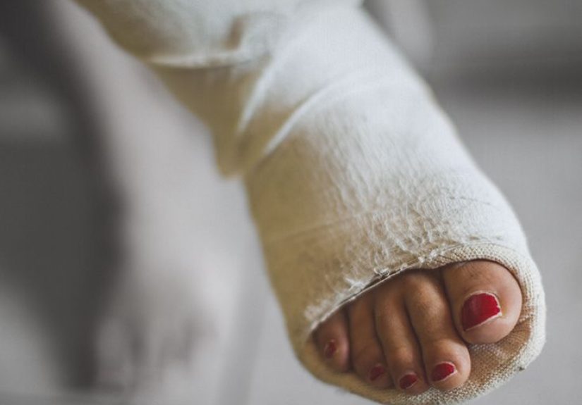
- Long bone fractures (especially femur, tibia) and pelvic fractures
- Multiple fractures or high-energy trauma (more marrow disruption, more opportunity for fat to enter veins)
- Orthopedic procedures that manipulate the medullary canal (for example intramedullary nailing/reaming)
- Joint replacement surgery (hip/knee arthroplasty) in certain settings
Less common triggers (the “surprise cameos”)
FES can also occur without classic fracture trauma. Reported non-traumatic or mixed triggers include severe burns,
pancreatitis, sickle cell–related marrow injury, bone marrow transplantation, and some cosmetic procedures (notably
liposuction or fat grafting). Certain medication/infusion contexts (like lipid emulsions) have also been discussed.
Who is at higher risk?
Risk rises with factors like multiple injuries, major fractures managed conservatively for longer periods, and
higher injury severity overall. Clinicians also watch more carefully when there are bilateral femur fractures,
associated thoraco-abdominal trauma, or hemodynamic instabilitybecause the overall physiologic stress is higher.
What’s going on inside the body? (The short science)
FES is often explained with two overlapping theories, and in real life it’s probably a messy duet:
1) Mechanical theory: “Fat droplets block tiny vessels”
Trauma or surgical manipulation can increase pressure inside the bone’s medullary canal, pushing fat/marrow contents into
torn venous channels. Those droplets travel to the lungs and can obstruct small pulmonary vessels. In some cases,
droplets (or inflammatory downstream effects) can also affect the brain and other organs.
2) Biochemical/inflammatory theory: “The body reacts… loudly”
Fat emboli aren’t just passive blobs. They can trigger inflammation and lead to release of free fatty acids and other
mediators that irritate the lining of blood vessels and lung tissue. The end result can look a lot like
acute respiratory distress syndrome (ARDS)which is ICU-speak for “the lungs are inflamed and oxygen exchange is struggling.”
Translation: FES is not only about blockage. It’s also about inflammation. That’s part of why symptoms can progress
and why supportive critical care matters.
Symptoms: the classic triad (plus the supporting cast)
Clinicians often talk about a “classic triad.” Not every patient gets all three, but it’s a famous combo:
- Respiratory symptoms: shortness of breath, fast breathing, low oxygen levels
- Neurologic changes: confusion, agitation, headache, seizures, reduced responsiveness
- Petechial rash: tiny red-purple spots (often on the chest, neck, axillae, conjunctiva, or oral mucosa)
Other possible findings include fever, fast heart rate, jaundice, anemia, low platelets, vision changes, and
signs of kidney stress. The rash is especially helpful when it appearsit’s like the body waving a small
dotted flag that says, “Hey, something systemic is happening.”
When is it an emergency?
If someone develops new breathing difficulty, worsening oxygen levels, or
new confusion after a major fracture or relevant surgery, they should be evaluated urgently.
FES can range from mild to life-threatening, and early supportive care improves safety.
Diagnosis: how doctors put the puzzle together
There’s no universally accepted single test that “proves” fat embolism syndrome on the spot.
Diagnosis is usually clinicalbuilt from risk factors, timing, exam findings, and tests that support the diagnosis
while ruling out other dangerous look-alikes (like pulmonary embolism, stroke, pneumonia, or lung contusion).
Clinical criteria you may hear about
Several diagnostic frameworks exist. The most commonly referenced include:
-
Gurd/Wilson criteria: uses a mix of “major” and “minor” features (petechiae, respiratory insufficiency,
cerebral involvement, fever, tachycardia, anemia, thrombocytopenia, and more). -
Schonfeld score: assigns points for findings like petechiae, diffuse chest infiltrates, hypoxemia,
fever, tachycardia, confusion. - Lindeque criteria: focuses more narrowly on respiratory parameters.
None of these is perfect. They’re best thought of as “structured ways to think,” not magical sorting hats.
Tests that often help
- Pulse oximetry / blood gases to document low oxygen
- Blood tests that may show anemia, low platelets, inflammation markers, or other changes
- Chest X-ray or CT to look for lung infiltrates and help rule out alternatives
- Brain imaging (CT/MRI) if neurologic symptoms occurMRI can be more sensitive for certain patterns
- Workup for “the mimics”: doctors often rule out blood-clot pulmonary embolism or stroke when symptoms overlap
Importantly, clinicians don’t diagnose FES just because fat droplets existfat can circulate after fractures without causing
the full syndrome. The diagnosis is about the clinical illness.
A real-world example (why teams watch closely)
In trauma literature, there are cases where a patient with multiple long-bone fractures develops acute hypoxemia,
and then neurologic changes. Rarely, fat emboli have been visualized in transit in large veins on imaging.
One reported case involved a patient with bilateral femur fractures who deteriorated and was later found to have
a patent foramen ovale (a heart connection that can allow emboli to cross from venous to arterial circulation),
complicating the neurologic picture. The big takeaway is not “panic,” but “stay vigilant and stabilize fractures promptly.”
Treatment: what actually happens (supportive care is the star)
Here’s the frustrating truth and the reassuring truth rolled into one:
there’s no single “cure” that instantly dissolves fat embolism syndrome.
Management focuses on supporting breathing and circulation, protecting organs while the body clears the insult,
and preventing additional embolic load (often by stabilizing fractures).
Supportive care: the main course
- Oxygen: from nasal cannula up to high-level support depending on severity
- Ventilation: if respiratory failure develops, intubation and mechanical ventilation may be needed
- Hemodynamic support: maintaining blood pressure and perfusion; treating shock if present
- Fluids and nutrition: enough to support recovery, without drowning the lungs
- DVT prevention: because trauma and immobilization can also raise blood-clot risk
Some protocols include albumin in resuscitation for certain patients because it helps restore intravascular volume
and may bind free fatty acidsone of the proposed inflammatory contributors.
Medications and interventions: what’s debated (and why)
-
Corticosteroids: sometimes used to reduce inflammation and support lung function. Evidence is mixed,
and some analyses suggest benefit in prevention in certain fracture patients, but routine use remains controversial. -
Heparin: once explored because it affects lipid metabolism, but it’s generally not used as a specific FES treatment
due to bleeding risk and lack of proven clinical benefit for FES itself. - IVC filters: proposed by some as a preventive strategy, but not well established specifically for fat emboli.
- ECMO: in rare, severe cases with profound respiratory failure, advanced support like ECMO may be considered.
Fracture stabilization: prevention and part of management
Early stabilization of long-bone fractures is widely viewed as a key prevention strategy and may reduce the incidence
of FES compared with prolonged conservative management. During orthopedic fixation, teams try to avoid unnecessarily high
intramedullary pressures, though specific techniques vary and not all have proven outcome benefits.
Recovery and outlook
Many casesespecially mild to moderateresolve over days with supportive care. Severe cases can be dangerous, particularly
when oxygen levels drop significantly or the heart struggles under increased strain from lung injury.
Reported mortality rates vary across studies and settings; modern care and prevention strategies have generally improved outcomes.
Some patients, especially those with significant neurologic involvement or severe ARDS-like lung injury, may need longer rehabilitation.
Prevention: what reduces risk?
- Prompt fracture stabilization when appropriate
- Careful perioperative technique during intramedullary instrumentation
- Close monitoring of high-risk trauma patients in the first 24–72 hours
- Thoughtful consideration of steroids in select contexts (still debated; not universally standard)
Frequently asked questions
Is fat embolism syndrome the same as a blood-clot pulmonary embolism?
No. A classic pulmonary embolism is usually a blood clot that traveled to the lungs. In FES, the embolic material is fat (often marrow fat),
and the syndrome also involves inflammation and multi-organ effects. The symptoms can overlap, which is why clinicians often rule out clots.
Does everyone with a long bone fracture get FES?
No. Fat droplets in the bloodstream can occur after fractures, but fat embolism syndrome is much less common.
Risk increases with multiple fractures, higher-energy trauma, and certain surgical or medical contexts.
What’s the most recognizable sign?
The petechial rash is distinctive when it appears, but it doesn’t happen in every case. Often the earliest major clue is worsening oxygenation
within a day or two after the inciting event.
How long does recovery take?
Mild cases may improve within days. More severe lung or neurologic involvement can take longer, sometimes requiring rehabilitation. Many patients
recover well with proper supportive care.
Conclusion
Fat embolism syndrome is a rare but serious complication most often associated with long bone or pelvic fractures and certain orthopedic procedures.
It typically appears within a few days and is recognized by a combination of respiratory distress, neurologic changes, and sometimes a telltale petechial rash.
Because there’s no single definitive test, timely recognition and supportive careespecially oxygenation and hemodynamic stabilityare the backbone of treatment,
while early fracture stabilization helps reduce risk.
Experiences related to fat embolism syndrome (what it can feel like, and what the hospital course often looks like)
People who go through a suspected or confirmed fat embolism syndrome episode often describe it as “the complication I didn’t know existed.”
The story frequently starts with a major injurylike a femur fracture from a car crash or a bad fallfollowed by a period where things seem stable.
Then, within the next day or two, breathing becomes noticeably harder. Some patients recall feeling suddenly winded while lying still, like their lungs
forgot the plan. Others don’t remember the onset at all because confusion or drowsiness can creep in before they have time to narrate the experience.
In the emergency department or ICU, the experience can feel oddly fast and slow at the same time. Fast: alarms, oxygen devices, more staff in the room,
urgent imaging, repeated vital signs, and questions that sound repetitive (“Any chest pain? Any new confusion?”). Slow: long hours watching monitors and
waiting for oxygen levels to settle. Families often notice the mental changes firstirritability, spacing out, or the patient seeming “not quite themselves.”
Clinicians take that seriously because neurologic symptoms can look like many other emergencies, so brain and lung imaging may be ordered to rule out stroke,
blood-clot pulmonary embolism, pneumonia, or lung contusion.
If a rash appears, it can be a weirdly validating momentnobody wants a rash, but petechiae can help clinicians connect the dots. Patients might notice
tiny red spots on the chest or around the eyes; clinicians may check the mouth and eyelids too. Lab draws become frequent, partly to monitor oxygenation
and organ function, and partly because FES can be accompanied by anemia or low platelets. Many patients describe the bloodwork schedule as “my new full-time job,”
which is a fair critique.
Treatment feels supportive because it is. People often remember oxygen as the centerpiecestarting with a nasal cannula and sometimes escalating.
If breathing worsens significantly, sedation and a ventilator can enter the picture. That can be scary for families, but for many patients it functions like
a temporary bridge while lung inflammation calms down. Meanwhile, orthopedic stabilizationsplinting, traction, or surgerymay happen sooner rather than later.
This can surprise patients (“Wait, surgery now?”), but early stabilization is often part of reducing ongoing embolic burden and improving overall recovery.
Recovery experiences vary. In mild cases, people report that oxygen needs fade over days and the mental fog lifts gradually.
In more severe cases, the after-effects can include fatigue, shortness of breath with exertion, and frustration at how long it takes to feel normal again.
Physical therapy becomes central: not only for the fracture, but for rebuilding stamina after a respiratory hit. Emotionally, many patients describe a
“double recovery”healing the bone and regaining confidence in breathing and cognition. A common theme is gratitude for early recognition and monitoring:
FES is unpredictable, but when teams anticipate it, patients often feel safereven if they wish the body’s bone marrow would stop freelancing.