
Table of Contents >> Show >> Hide
- What Is a Pregnancy Ultrasound, Exactly?
- Why Pregnancy Ultrasounds Are Done
- Types of Pregnancy Ultrasound
- How to Prepare for a Pregnancy Ultrasound
- Step-by-Step: What Happens During a Pregnancy Ultrasound
- Is Pregnancy Ultrasound Safe?
- Understanding Your Ultrasound Results
- Smart Questions to Ask Your Provider
- Real-Life Pregnancy Ultrasound Experiences & Tips
- Bottom Line
Few things change your relationship with your body like pregnancy. One day you’re just you; the next, there’s a tiny roommate whose favorite hobbies include growing organs and kicking your bladder. Pregnancy ultrasound is the main way your care team checks in on that little roommate and makes sure the house (your uterus) is staying in good shape. Think of it as a high-tech, completely sound-wave–powered window into what’s going on inside.
What Is a Pregnancy Ultrasound, Exactly?
A pregnancy ultrasound (also called a prenatal ultrasound, fetal ultrasound, or obstetric ultrasound) is an imaging test that uses high-frequency sound waves, not radiation, to create pictures of your uterus, ovaries, placenta, amniotic fluid, and baby. A handheld device called a transducer sends sound waves into the body and listens for the echoes, which a computer turns into images in real time.
Unlike X-rays or CT scans, obstetric ultrasound does not use ionizing radiation and has no known harmful effects when it’s done for medical reasons by trained professionals. That’s why major organizations consider it the preferred way to monitor pregnancy.
Why Pregnancy Ultrasounds Are Done
Pregnancy ultrasounds have different jobs at different stages of pregnancy. Your provider may not order every possible scan, but here’s what they’re generally looking for over the course of nine months.
Early Pregnancy (Around 6–10 Weeks)
- Confirm that you’re pregnant and that the embryo is in the uterus (not ectopic).
- Check for a heartbeat and basic viability.
- Measure the embryo to estimate how far along you are and calculate your due date.
- See whether you’re carrying one baby, twins, or more.
- Investigate symptoms like bleeding or significant pain.
These early ultrasounds are especially helpful if you’re unsure of your last menstrual period date or you’ve had prior complications like miscarriage or ectopic pregnancy.
First Trimester Dating or Nuchal Translucency Scan (Around 11–13 Weeks)
- Confirm gestational age and refine your estimated due date.
- Check baby’s overall growth, early anatomy, and position in the uterus.
- Verify how many babies you’re carrying.
- Measure the fluid at the back of baby’s neck (nuchal translucency) as part of first-trimester screening for some chromosome conditions.
Second Trimester Anatomy Scan (Typically 18–22 Weeks)
This is the “big” ultrasound many people think ofthe detailed anatomy scan. It’s like a full-body checkup for your baby on screen. Your provider or sonographer will:
- Measure baby’s head, abdomen, and limbs to assess growth.
- Look closely at brain, spine, heart, kidneys, stomach, bladder, face, limbs, and more.
- Check the placenta’s location, umbilical cord, and amniotic fluid.
- Estimate due date and fetal weight range.
- Tell you the baby’s sex, if you want to know and the position cooperates.
A standard second-trimester ultrasound is designed to screen for major structural differences or anomalies and to confirm that baby is growing as expected.
Third Trimester and High-Risk Pregnancy Ultrasounds
Not everyone has a third-trimester ultrasound, but your provider may order one if they need to:
- Check baby’s growth, especially if you’re measuring small or large for dates.
- Evaluate amniotic fluid levels.
- Confirm baby’s position (head-down, breech, transverse).
- Monitor the placenta if there have been concerns (such as placenta previa or growth restriction).
- Track twins or multiples or complications like gestational diabetes or high blood pressure.
In high-risk pregnancies, you might also have specialized ultrasoundslike Doppler studies to measure blood flow or additional growth scans to make sure baby and placenta are doing well.
Types of Pregnancy Ultrasound
The term “pregnancy ultrasound” covers several different techniques. Your care team will choose the one that gives them the clearest view with the least hassle for you.
Transabdominal Ultrasound
This is the classic scenario you’ve seen in movies: warm gel on your belly, transducer gliding around, baby on the screen. A transabdominal ultrasound is common in the second and third trimester and often later in the first trimester once the uterus has risen out of the pelvis.
Transvaginal Ultrasound
Early in pregnancy, the uterus sits low in the pelvis, so the clearest pictures often come from a transvaginal ultrasound. The sonographer places a narrow, covered, lubricated probe a short distance into the vagina to get closer to the uterus. It shouldn’t be painful, but it can feel a bit strange or intimate; you’ll typically be draped and can request a support person or chaperone if you’d like.
Doppler Ultrasound
Doppler ultrasound looks at blood flowthrough the umbilical cord, placenta, or specific fetal vessels. It’s often used later in pregnancy or in higher-risk situations to make sure oxygen and nutrients are getting where they need to go.
2D vs. 3D and 4D Ultrasound
Most medical scans are 2D (two-dimensional), which is more than enough to check growth and anatomy. Some centers also offer 3D (still) or 4D (moving) images that give more lifelike views of the face or limbs. While these can be amazing keepsakes, leading groups and the U.S. FDA caution against “keepsake” ultrasound sessions done outside a medical setting just for entertainment, since they can mean longer, unnecessary exposure by untrained operators.
How to Prepare for a Pregnancy Ultrasound
The good news: prep is usually simple. No week-long juice cleanse, no special supplements, and you definitely don’t need to shave your belly (unless you just feel like it).
Before an Early or First-Trimester Ultrasound
- Ask about your bladder. Many clinics ask you to arrive with a comfortably full bladder for early or dating scans. The full bladder pushes the uterus into a better position and improves the clarity of the images.
- Take your usual medicines. Unless your provider says otherwise, continue routine medications.
- Wear a two-piece outfit. It’s easier to expose your abdomen without fully undressing.
- Bring paperwork and questions. Insurance card, ID, previous imaging reports if you have them, and a list of anything you want to ask.
Before a Second or Third Trimester Ultrasound
- You can typically eat and drink normally beforehand.
- Some centers still prefer a partially full bladder for anatomy scans; others don’t. Follow the specific instructions you receive.
- Avoid heavy lotions or oils on your belly the day of the scan; these can interfere with the gel making good contact.
- Ask about guest policies. Many clinics allow one or two support people but may limit children so the sonographer can focus.
What to Bring (Emotionally, Not Just in Your Bag)
- A realistic mindset: most ultrasounds are reassuring, but sometimes they find things that need follow-up.
- A support person if that makes you feel safer or more relaxed.
- Your curiositythis is your visit and your baby. It’s okay to ask, “What are we looking at now?”
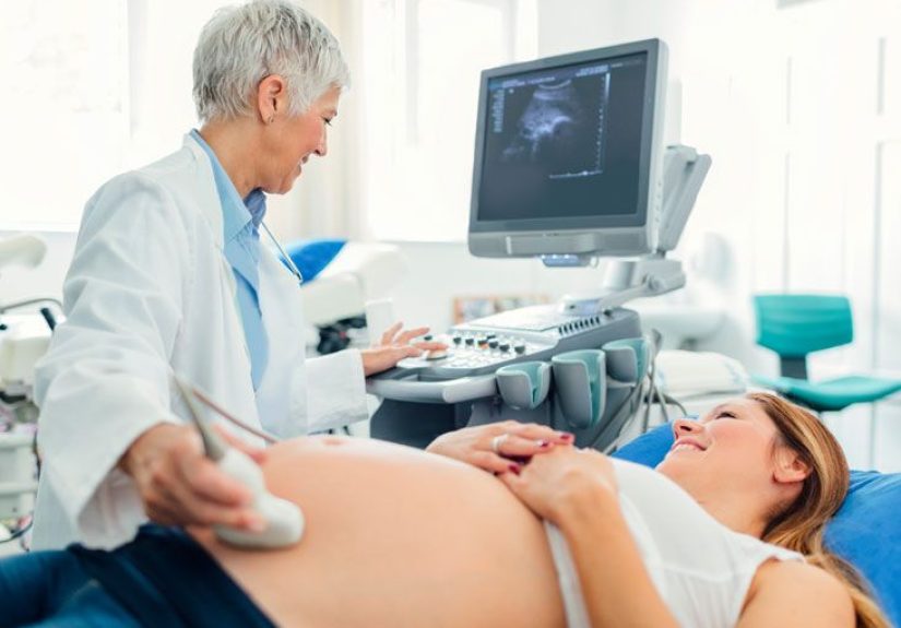
Step-by-Step: What Happens During a Pregnancy Ultrasound
While every clinic has its own flow, the basic procedure is pretty consistent. Here’s how it usually goes for a transabdominal scan:
- Check-in. You’ll confirm your identity, due date (if known), and the reason for the scan.
- Getting ready. You’ll lie on an exam table, usually on your back or slightly tilted to one side. The sonographer may tuck a towel into your waistband to keep gel off your clothes.
- Gel time. Warm (or occasionally not-quite-warm) gel is applied to your abdomen. It helps the transducer glide and improves sound-wave transmission.
- Imaging. The sonographer moves the transducer over your belly, sometimes pressing more firmly to get certain angles. You’ll see gray-scale images on the screen in real time.
- Measurements and screenshots. They’ll freeze images to measure baby’s head, abdomen, limbs, and other structures, and capture short clips for your provider to review.
- Listening to the heartbeat. When it’s appropriate, you may hear your baby’s heartbeatoften a highlight of the visit.
- Review and discussion. Depending on the clinic, the sonographer may explain basics as they go, or you may speak with your obstetric provider afterward to go over results.
For a transvaginal ultrasound, the steps are similar, but the transducer is gently inserted into the vagina instead of placed on your abdomen. The exam usually lasts 15–45 minutes, depending on what your team needs to evaluate.
Is Pregnancy Ultrasound Safe?
The short answer: when medically indicated and performed by trained professionals, yes. Ultrasound has been used in pregnancy for decades, and major organizations agree that it has no known harmful effects at the energy levels used clinically.
A few important safety takeaways:
- No ionizing radiation. Ultrasound is different from X-rays and CT scans, which involve radiation exposure. That’s one reason it’s the go-to imaging method during pregnancy.
- Use it when you need it. Professional groups recommend that ultrasounds be performed when there’s a medical reasonnot every week just for fun.
- Skip non-medical “keepsake” sessions. The FDA and professional societies warn against commercial studios that offer long, entertainment-only ultrasounds. More time on the machine doesn’t mean better pictures, and untrained operators may not follow safety standards.
Understanding Your Ultrasound Results
After the scan, the sonographer and/or radiologist will interpret the images and send a report to your obstetric provider. You might get a quick “Everything looks great!” on the spot, or you might need a follow-up visit to discuss details.
Results might include:
- Gestational age and due date. Based on measurements, especially in early pregnancy.
- Fetal growth. Is baby measuring appropriately for the gestational age?
- Fetal anatomy. Structures like the brain, heart, spine, kidneys, and limbs are assessed for typical development.
- Placenta and umbilical cord. Location, attachment, and blood flow.
- Amniotic fluid. Too much or too little fluid can signal a need for closer monitoring.
- Number and position of babies. Single or multiple pregnancy, breech or head-down, and so on.
If the ultrasound shows something unexpected, it doesn’t automatically mean there’s a serious problem. Sometimes baby’s position or body size just makes things harder to see, so your provider may recommend a repeat scan, a detailed anatomy ultrasound, or a referral to a maternal-fetal medicine specialist for a closer look.
Smart Questions to Ask Your Provider
Want to get the most out of your pregnancy ultrasound? Consider asking:
- “What’s the main reason for this ultrasound today?”
- “How far along am I based on today’s measurements?”
- “Are the baby’s growth and fluid levels in the expected range?”
- “Did you see anything that needs follow-up or extra testing?”
- “If I’m high-risk, how often might I need ultrasounds?”
- “Is there anything I should watch for at home between now and my next visit?”
Real-Life Pregnancy Ultrasound Experiences & Tips
On paper, pregnancy ultrasounds sound very clinical: sound waves, measurements, biometric data. In real life, they’re often emotional milestonestiny movie premieres where your baby is both the director and the star.
Many people say the first time they hear the heartbeat, everything suddenly feels very real. Before that, pregnancy can feel like a mix of nausea, exhaustion, and a stack of appointment cards. Seeing that little flicker and hearing the rapid whoosh-whoosh of the fetal heart often shifts things from “Is this really happening?” to “Oh wow, this is happening.”
At early scans, don’t be surprised if baby looks more like a lima bean with a blinking light than the serene babies you see in social media posts. That’s normal. By the time you reach the anatomy scan, the images are often much more detailedtiny fingers, a profile shot, maybe even a well-timed kick that makes the whole exam room laugh.
It’s also normal to feel nervous before an ultrasound, especially if you’ve had pregnancy complications in the past or waited a long time to conceive. One helpful strategy is to separate the appointment into two parts in your mind: the information (what your team needs to see) and the moment (your experience of seeing your baby). Bringing a support person who understands that can make a big differencethey can ask practical questions while you just breathe and watch.
If you’re the type who copes with stress by preparing, consider journaling your questions the day before the scan. You might jot down things like “Ask whether baby’s growth is on track” or “Clarify what they’re looking for in the placenta.” After the appointment, you can add a short note about how it went and what you sawover time, those notes become a kind of pregnancy diary you may really treasure.
Another tip: manage your expectations about the pictures you’ll take home. Some babies pose like they’ve read the ultrasound manualperfect profile, tiny hand up for a wave. Others tuck into your pelvis, face the spine, or keep their hands over their face. If your images look more like abstract art than baby photos, it doesn’t mean anything is wrong; it just means your baby is already a free spirit with strong opinions about paparazzi.
And yes, it’s okay to feel a little overwhelmed afterward, even when the news is good. You’ve just absorbed a lot: medical information, emotional reactions, and a visual reminder that there’s a new human being growing inside you. Some people celebrate with a favorite snack or a quiet walk; others send ultrasound pictures to their entire contact list within minutes. There’s no “right” way to feelonly the way that helps you move through this experience with as much calm and self-compassion as possible.
The most important takeaway? A pregnancy ultrasound isn’t just a box to check; it’s one of the main tools your care team uses to keep both you and your baby as safe and healthy as possible. Understanding the purpose, procedure, and preparation helps turn the appointment from something you simply “get through” into something you actively participate inwith questions, curiosity, and maybe a happy tear or two.
Bottom Line
Pregnancy ultrasounds are a cornerstone of prenatal care, providing vital information about your baby’s health, your uterus and placenta, and the overall progress of your pregnancy. When used appropriately, they’re safe, noninvasive, and incredibly informative. Knowing what to expectand how to preparecan make each scan less stressful and more meaningful, whether it’s your very first fuzzy image or the final check before delivery.