
Table of Contents >> Show >> Hide
- Quick Answer: Yes, But Usually Not in the Way People Think
- Why Lesions Matter So Much in MS Diagnosis
- So Why Might MS Be Suspected Even If Brain and Spine Scans Look Clean?
- Can You Be Diagnosed With MS Without Visible Brain and Spinal Lesions?
- What Doctors Do When MS Is Suspected but MRI Looks Normal
- Conditions That Can Look Like “Lesion-Free MS”
- What This Means for Patients
- Experiences Related to “Can You Have MS Without Brain and Spinal Lesions?”
- Conclusion
Here’s the short version: yes, sometimes a person can have symptoms that strongly suggest multiple sclerosis even when standard brain and spinal MRIs do not show the classic lesions doctors expect. But that answer needs a giant asterisk. In real-life neurology, MS is usually diagnosed by finding evidence of damage in the central nervous system over time, often with MRI support. So if both the brain and spinal cord look completely normal on a high-quality scan, especially on repeated scans, doctors usually become much more cautious about calling it MS.
In other words, a clean MRI does not automatically slam the door on MS, but it does make neurologists pause, reread the clues, and ask whether another condition is trying to sneak into the room wearing an MS costume. Sometimes the explanation is that lesions are in a place routine imaging missed. Sometimes the problem is that it’s simply too early. And sometimes the truth is that the symptoms come from something entirely different.
Quick Answer: Yes, But Usually Not in the Way People Think
When people ask, “Can you have MS without brain and spinal lesions?” they usually mean one of three things:
1. A normal first MRI
This can happen. Someone may have symptoms such as numbness, weakness, vision changes, balance problems, or electric-shock sensations, yet their first scan looks normal or nonspecific. That does not prove they have MS, but it also does not always rule it out on day one.
2. No visible lesions on routine imaging, but disease is elsewhere
MS can affect the optic nerve, and optic nerve involvement does not always jump out on a standard brain MRI. In some cases, doctors need orbital MRI, optical coherence tomography, visual evoked potentials, or spinal fluid testing to get the full picture. So a person may seem “lesion-free” on a routine brain-and-spine workup while still having objective evidence of demyelination elsewhere.
3. Repeatedly normal brain and spine MRI over time
This is where the story changes. If a patient has long-standing neurologic symptoms and repeated, high-quality MRIs of the brain and spinal cord remain normal, classic MS becomes much less likely. At that point, neurologists often take a harder look at other diagnoses, because MS is a diagnosis of evidence, not wishful thinking.
Why Lesions Matter So Much in MS Diagnosis
Multiple sclerosis is a disease of the central nervous system, which includes the brain, spinal cord, and optic nerves. The immune system mistakenly attacks myelin, the protective coating around nerve fibers, and that damage can leave scars or lesions. Those lesions are not just decorative drama on a radiology report. They help doctors prove that inflammation has occurred in more than one place and, in many cases, at more than one time.
That is why MRI is such a big deal in MS. It helps confirm the diagnosis, supports treatment decisions, tracks disease activity, and helps rule out impostors like neuromyelitis optica spectrum disorder, MOG antibody disease, migraine-related white matter changes, vitamin deficiencies, strokes, infections, and spinal compression. MRI is powerful. It is just not magic. It cannot read minds, predict the future, or explain every symptom after one scan and a cup of coffee.
Doctors do not diagnose MS from one symptom alone. They combine medical history, neurologic exam, MRI findings, spinal fluid results, and sometimes vision testing or repeat imaging. The goal is not just to diagnose fast. It is to diagnose correctly.
So Why Might MS Be Suspected Even If Brain and Spine Scans Look Clean?
The MRI may not have been good enough
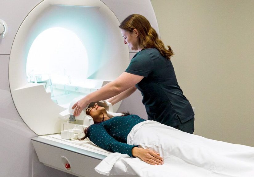
Not all MRIs are created equal. A low-quality study, motion-blurred scan, limited sequence set, or imaging done on a weaker machine can miss subtle lesions. Spinal cord imaging is especially tricky because the cord is narrow, moves with breathing and heartbeat, and is more prone to artifact. That is why MS specialists often prefer dedicated protocols and good magnet strength when the diagnosis is on the line.
The lesions may be in the optic nerve
One of the biggest updates in modern MS thinking is the growing recognition that the optic nerve matters. A person may have optic neuritis, pain with eye movement, blurred vision, or reduced color vision, yet routine brain and spine imaging may not fully capture the problem. In those cases, orbital MRI, OCT, and visual testing can reveal evidence that standard imaging missed.
The disease may be early
Sometimes symptoms arrive before the classic lesion pattern becomes obvious. A person may experience a clinically isolated syndrome, which is a first neurologic episode suspicious for demyelination, but not yet enough to prove MS. Over time, repeat MRI or spinal fluid testing may clarify whether this was the opening chapter of MS or a one-time event with a completely different explanation.
Conventional MRI can miss cortical damage
MS is often described as a white matter disease, but it can also affect gray matter, including the cortex. Conventional MRI is better at spotting some lesions than others, and cortical lesions are notoriously easy to miss. Higher-field imaging and advanced techniques can detect more of them. That means a scan that looks “quiet” may not always mean the nervous system is perfectly untouched.
Rare disease patterns can complicate the picture
Research has described unusual patterns of MS pathology, including forms with less obvious brain white matter involvement than expected. These are not the everyday version of MS, and they do not mean doctors should diagnose MS casually when imaging is normal. What they do mean is that neurology still has some plot twists left in the script.
Can You Be Diagnosed With MS Without Visible Brain and Spinal Lesions?
Usually, doctors want objective evidence somewhere in the central nervous system. Historically, that often meant lesions in characteristic locations on brain or spinal MRI. Today, the conversation is a little more nuanced. The optic nerve can count. Cerebrospinal fluid can help. Certain biomarkers and newer imaging features can strengthen the case.
But here is the key distinction: being diagnosed without visible brain and spinal lesions on a routine scan is not the same as being diagnosed with no objective evidence of disease at all. If there is no supportive MRI finding, no optic nerve evidence, no spinal fluid abnormality, no clear clinical attacks, and no other objective marker, neurologists generally become very hesitant to label the problem as MS.
That hesitation is a good thing. MS treatments can be life-changing, but they are not casual vitamins from a checkout aisle. A mistaken diagnosis can expose someone to years of unnecessary medication, side effects, anxiety, insurance headaches, and a completely wrong explanation for their symptoms.
What Doctors Do When MS Is Suspected but MRI Looks Normal
Repeat imaging
If the symptoms are truly suspicious, doctors may repeat MRI after a period of time. MS can declare itself later, and new lesions may appear on follow-up scans even when the first imaging was unrevealing.
Use better-targeted imaging
This may include dedicated cervical and thoracic spine MRI, orbital MRI for suspected optic neuritis, or imaging at a center with MS-focused protocols. Sometimes the issue is not that there are no lesions. It is that the original scan was not asking the right question in the right way.
Order a lumbar puncture
Spinal fluid can show oligoclonal bands or other immune activity that supports MS. These findings are not perfect and do not diagnose MS by themselves, but they can be very helpful when the clinical picture is strong and MRI evidence is incomplete.
Check for mimics
Blood tests and targeted workups may look for vitamin B12 deficiency, thyroid disease, lupus, Sjögren’s syndrome, sarcoidosis, infections, NMOSD, MOGAD, or other inflammatory and structural causes. This part of the workup is not a side quest. It is central to getting the diagnosis right.
Re-examine the symptoms themselves
Neurologists pay close attention to whether symptoms fit a typical demyelinating pattern. For example, vague tingling all over the body, non-anatomical sensory complaints, or chronic fatigue without objective neurologic findings may prompt a broader search instead of a fast MS label.
Conditions That Can Look Like “Lesion-Free MS”
Several disorders can mimic MS, especially early on:
Migraine and small vessel changes
White matter spots from migraine or aging blood vessels can confuse the picture, especially if someone has headaches, dizziness, or sensory symptoms. These spots are not automatically MS, even if the MRI report sounds dramatic.
NMOSD
Neuromyelitis optica spectrum disorder can affect the optic nerves and spinal cord and may look like MS at first glance. But it is a different disease with different treatment implications. Early in its course, the brain MRI may even be relatively normal.
MOG antibody disease
MOGAD can cause optic neuritis, myelitis, or brain inflammation and is another reason doctors should avoid making an MS diagnosis too quickly. MRI patterns and antibody testing can help separate it from MS.
Vitamin deficiency or metabolic causes
Vitamin B12 deficiency, copper deficiency, thyroid disorders, and other metabolic problems can trigger numbness, weakness, imbalance, and cognitive complaints that overlap with MS symptoms.
Structural spine disease
Cervical spondylosis, disc disease, and spinal cord compression can produce numbness, weakness, gait issues, and electric-shock sensations. Sometimes the spine, not the immune system, is the real culprit.
Functional neurologic disorder and other non-demyelinating causes
Some symptoms are very real but do not come from inflammatory lesions in the central nervous system. That does not mean “nothing is wrong.” It means the right diagnosis may live outside the MS universe.
What This Means for Patients
If you have symptoms but no brain or spinal lesions, the next best step is not panic and definitely not self-diagnosing through a 2 a.m. search spiral. The better move is a careful evaluation by a neurologist, ideally one familiar with MS and its mimics. Ask whether the MRI quality was adequate, whether the optic nerves were evaluated, whether spinal fluid testing is needed, and whether other causes have been meaningfully excluded.
If your scans remain normal over time, that does not mean your symptoms are imaginary. It means the explanation may be something other than MS, and finding that explanation matters just as much as ruling MS in or out. A good neurologist is not trying to win a speed round. They are trying to get the map right before anyone starts driving.
Experiences Related to “Can You Have MS Without Brain and Spinal Lesions?”
One of the most frustrating parts of this topic is that the patient experience often feels messier than the textbook version. Many people do not walk into a clinic saying, “Hello doctor, I have a perfectly organized demyelinating episode that will line up beautifully with diagnostic criteria.” They come in saying their foot went numb for a week, their vision got weird for three days, or their legs feel heavy even though their first MRI report says something unhelpful like “no acute abnormality.” That gap between symptoms and proof can feel enormous.
Some people spend months in diagnostic limbo after a normal or inconclusive brain MRI. They know something is off, but they do not yet have the radiology evidence to match the feeling. In those cases, the experience is often a mix of relief and irritation. Relief, because nobody wants scary lesions on a scan. Irritation, because a normal test does not erase a very real symptom. For many patients, the hardest part is not pain or numbness. It is uncertainty.
Others have the opposite experience. They are initially told the MRI is “probably fine,” then later a specialist reviews the images and notices that the study did not include the right sequences, did not adequately image the spinal cord, or never looked carefully at the optic nerves. Suddenly the story changes from “maybe nothing” to “we need a repeat scan with an MS protocol.” That shift can be emotionally exhausting. People often feel as if the floor keeps moving under them.
There are also patients whose symptoms turn out not to be MS at all, and their stories matter just as much. Someone may arrive convinced they have lesion-free MS, only to learn they actually have migraine with sensory aura, MOG antibody disease, vitamin B12 deficiency, cervical myelopathy, autoimmune disease, or a completely different neurologic disorder. That can be disappointing in a strange way. After weeks or months of focusing on one feared diagnosis, the brain gets attached to the theory. Letting go of it is not always easy, even when the new answer fits better.
Then there are people whose diagnosis becomes clear only with time. Their first scan is normal. Their second scan is not. Or their MRI stays unclear, but spinal fluid, orbital imaging, and the pattern of attacks finally make the picture coherent. These patients often describe the diagnosis as both upsetting and validating. Upsetting, because MS is a life-changing condition. Validating, because the mystery finally has a name.
The common thread in all these experiences is that MS diagnosis is rarely just a medical event. It is also a psychological journey through uncertainty, second opinions, repeat testing, and a lot of waiting-room ceiling tiles. That is why clear communication matters so much. Patients do better when clinicians explain that “normal MRI” does not always mean “case closed,” but also that it should prompt a serious search for alternatives. The most useful message is usually the most honest one: we do not know everything from one scan, but we do know enough to keep looking carefully and avoid jumping to the wrong conclusion.
Conclusion
So, can you have MS without brain and spinal lesions? In a limited sense, yes. A person may have suspected MS even when standard brain and spine imaging is unrevealing, especially early on, when lesions are subtle, when the optic nerve is involved, or when routine MRI misses cortical disease. But if repeated, high-quality brain and spinal MRIs remain normal, classic MS becomes much less likely, and doctors should seriously consider other explanations.
The smartest answer is not a dramatic yes or no. It is this: MS without obvious brain and spinal lesions is possible at the edges of diagnosis, but MS without objective evidence somewhere in the central nervous system is a very hard diagnosis to defend. That is why the best workup is thorough, patient, and a little allergic to shortcuts.