
Table of Contents >> Show >> Hide
- What Is Dysdiadochokinesia?
- What Does It Feel Like in Real Life?
- How Doctors Diagnose Dysdiadochokinesia
- Common Causes of Dysdiadochokinesia
- Symptoms That Often Show Up Alongside It
- How Dysdiadochokinesia Is Treated
- Exercises and Rehab Strategies That May Help
- When to Seek Medical Attention Right Away
- What Recovery and Outlook Can Look Like
- Experiences Related to Dysdiadochokinesia: What Life Can Look Like
- Final Thoughts
Note: This article is for general educational purposes only and is not a substitute for medical diagnosis, emergency care, or personalized treatment from a licensed clinician.
Dysdiadochokinesia is one of those medical words that sounds like it was invented after someone spilled alphabet soup on a neurology textbook. But the meaning is surprisingly practical: it describes trouble performing rapid alternating movements, such as flipping the hand back and forth, tapping the foot quickly, or opening and closing the fist in a steady rhythm.
On its own, dysdiadochokinesia is not a disease. It is a clinical sign. In plain English, it is a clue. When a person cannot smoothly switch between opposite movements, clinicians start thinking about the cerebellum and the networks connected to it. That matters because the cerebellum helps coordinate timing, precision, balance, and the smooth handoff between one movement and the next. When that system is disrupted, the body may look awkward, delayed, shaky, or strangely out of sync.
This article breaks down what dysdiadochokinesia is, how it is diagnosed, what can cause it, which therapies and exercises may help, and what daily life can look like for people dealing with it. The goal is simple: make a complicated neurologic sign easier to understand without turning it into a boring textbook hostage situation.
What Is Dysdiadochokinesia?
Dysdiadochokinesia means an impaired ability to perform rapid alternating movements. A neurologist may ask a person to rapidly pronate and supinate the forearm, tap the fingers, open and close the hand, or tap the foot. If those movements are slow, irregular, poorly timed, or fall apart altogether, dysdiadochokinesia may be present.
It is most commonly associated with cerebellar dysfunction. The cerebellum sits toward the back of the brain and acts like a high-speed quality-control department for movement. It does not just help you move. It helps you move well. It fine-tunes force, timing, rhythm, and accuracy, and it helps coordinate opposing muscle groups so one movement can stop while another begins. When that timing system misfires, rapid alternating movement becomes especially difficult.
Dysdiadochokinesia often shows up alongside other cerebellar signs, including ataxia, dysmetria, slurred speech, gait imbalance, intention tremor, and abnormal eye movements. That is why clinicians rarely interpret it in isolation. It is part of a bigger neurologic picture.
What Does It Feel Like in Real Life?
Most people do not wake up and announce, “I appear to have dysdiadochokinesia.” They usually notice everyday problems first. A hand may feel clumsy while buttoning a shirt. Typing may become uneven. Fast alternating tasks like flipping a spatula, brushing teeth, twisting a doorknob, or tapping to a beat may feel strangely chaotic. Speech can also be affected, especially when quick, repeated syllables lose crisp timing.
Some people mainly notice arm and hand issues. Others struggle more with leg movements, walking, or balance. If the underlying cause is progressive, the changes may creep in gradually. If the cause is acute, such as stroke or bleeding, the symptoms can appear suddenly and dramatically.
How Doctors Diagnose Dysdiadochokinesia
1. The neurologic exam comes first
The diagnosis usually starts with a bedside neurologic exam. A clinician watches how well a person performs rapid alternating tasks. Common exam maneuvers include:
- Rapid forearm pronation and supination
- Fast finger tapping
- Opening and closing the fists quickly
- Foot tapping
- Speech-rate testing using repeated syllables
The clinician is not just checking speed. They are also watching rhythm, accuracy, fatigue, symmetry, and whether the movement breaks down over a few seconds. A person with dysdiadochokinesia may start reasonably well, then lose pattern, overshoot, hesitate, or produce sloppy, arrhythmic motion.
2. The exam rarely stops at one sign
Because dysdiadochokinesia usually points to broader neurologic dysfunction, clinicians also look for other abnormalities. These may include gait problems, wide-based walking, poor balance, dysmetria on finger-to-nose or heel-to-shin testing, slurred or scanning speech, tremor as the hand approaches a target, difficulty swallowing, or abnormal eye movements. The surrounding signs help narrow down whether the issue is likely cerebellar and how extensive it may be.
3. Finding the cause is the real diagnostic job
Once dysdiadochokinesia is identified, the next step is figuring out why. That workup often includes a medical history, medication review, family history, and a timeline of symptoms. Blood and urine studies may help look for vitamin deficiencies, metabolic problems, infection, inflammation, or toxic exposures. Brain imaging, especially MRI, may be used to identify stroke, bleeding, tumor, structural lesions, cerebellar degeneration, or multiple sclerosis. If hereditary ataxia is suspected, genetic testing may be part of the evaluation.
In short, the sign is diagnosed at the bedside, but the cause often requires deeper detective work.
Common Causes of Dysdiadochokinesia
Dysdiadochokinesia is linked to cerebellar dysfunction or damage to cerebellar pathways. Causes range from temporary and treatable to chronic and progressive. Common categories include:
Stroke and bleeding
Cerebellar stroke or intracranial hemorrhage can disrupt coordination suddenly. This is one reason abrupt onset should never be brushed off as “just clumsiness.”
Traumatic brain injury
Injury affecting the cerebellum or nearby pathways can impair rapid alternating movement, balance, and speech.
Multiple sclerosis and other inflammatory disorders
Inflammation in the central nervous system can interfere with cerebellar circuits and produce ataxia-like findings, including dysdiadochokinesia.
Neurodegenerative and hereditary ataxias
Conditions such as spinocerebellar ataxias, Friedreich ataxia, multiple system atrophy, and other degenerative disorders may cause progressive coordination problems over time.
Vitamin and metabolic problems
Deficiencies in vitamins such as B12, vitamin E, or thiamine can contribute to neurologic dysfunction. Metabolic or autoimmune conditions may also be involved.
Toxins, alcohol, and medications
Alcohol misuse, sedatives, certain medications, and toxic exposures can interfere with cerebellar function. Sometimes the cause is reversible once the trigger is identified and addressed.
Infections, tumors, and structural problems
Brain infections, abscesses, tumors, hydrocephalus, and other structural lesions can affect the cerebellum and change coordination.
The big takeaway is this: dysdiadochokinesia is not the finish line. It is the signpost pointing toward an underlying neurologic issue that needs attention.
Symptoms That Often Show Up Alongside It
Because dysdiadochokinesia is usually part of a larger cerebellar pattern, other symptoms often travel with it. These may include:
- Unsteady gait or a wide-based walk
- Poor balance and frequent near-falls
- Difficulty with fine motor tasks
- Slurred, slow, or choppy speech
- Tremor during movement
- Trouble reaching a target accurately
- Abnormal eye movements or visual tracking problems
- Difficulty swallowing in more advanced cases
Not every person will have every symptom, and the pattern varies depending on the underlying condition, how quickly it developed, and which parts of the nervous system are involved.
How Dysdiadochokinesia Is Treated
There is no single pill that “fixes” dysdiadochokinesia itself. Treatment focuses on the underlying cause and on improving function.
If the problem is due to a treatable condition, such as vitamin deficiency, immune-mediated disease, medication effect, toxic exposure, or infection, treating that cause may improve the coordination problem. In acute emergencies like stroke, hemorrhage, or expanding structural lesions, urgent medical care is essential.
For chronic cerebellar disorders, treatment often centers on rehabilitation and safety. That may include:
- Physical therapy for balance, gait, strength, and mobility
- Occupational therapy for hand function, daily tasks, and adaptive strategies
- Speech therapy for speech timing, articulation, swallowing, and communication
- Assistive devices such as canes, walkers, or other mobility aids
- Home-safety changes to reduce fall risk
In some cases, clinicians may also treat related symptoms such as tremor, spasticity, mood changes, or sleep problems. Progress depends heavily on the cause. Some people improve meaningfully. Others focus on slowing functional decline, staying safe, and preserving independence for as long as possible.
Exercises and Rehab Strategies That May Help
Exercises do not magically erase cerebellar dysfunction, but they can help improve control, maintain mobility, and support daily function. The smartest approach is a customized program created by a neurologist, neuro-physical therapist, occupational therapist, or speech-language pathologist. Quality matters more than speed. Sloppy reps are not heroics; they are just sloppy reps.
1. Rapid alternating movement drills
These target the very movement pattern that dysdiadochokinesia disrupts. A therapist may use:
- Forearm pronation and supination drills
- Finger tapping sequences
- Fast open-and-close hand movements
- Alternating toe or foot taps
The goal is usually controlled rhythm before speed. Many people do better when they begin slowly, use a metronome or verbal cue, and gradually increase pace only if movement quality holds up.
2. Targeted coordination practice
Because dysdiadochokinesia often overlaps with dysmetria and general incoordination, therapists may also use target-based tasks such as reaching to specific points, finger-to-target practice, or lower-extremity coordination tasks. These exercises can help build smoother transitions and better motor timing.
3. Balance and gait work
Core stability, balance training, safe transfers, and gait practice are often essential. Programs may include standing balance work, weight shifting, step training, walking drills, treadmill work in selected patients, and fall-prevention strategies. The aim is not just better clinic performance, but safer movement in the kitchen, bathroom, sidewalk, and life in general.
4. Strength and endurance training
Weakness is not always the primary problem in cerebellar dysfunction, but better strength and endurance can still support function. Therapists may incorporate lower-body strengthening, postural work, cycling, or structured aerobic activity when appropriate.
5. Speech-focused exercises
If speech is affected, a speech-language pathologist may work on alternate motion rate tasks, clarity, pacing, breath support, and compensatory strategies. That can make speech more intelligible and less exhausting.
6. Occupational therapy for real-world tasks
Occupational therapy is where “how do I live with this?” gets practical answers. Therapy may focus on dressing, handwriting, typing, eating, phone use, grooming, and safer strategies for cooking or bathing. Adaptive tools can make a major difference, especially when hand coordination is unreliable.
One important caveat: exercises should match the person’s cause, fall risk, and overall neurologic status. If symptoms are new, worsening quickly, or unexplained, do not start a random internet workout plan and hope for the best. Get evaluated first.
When to Seek Medical Attention Right Away
Sudden coordination problems deserve urgent evaluation, especially if they appear with headache, vomiting, facial droop, weakness, numbness, new speech changes, confusion, severe dizziness, or trouble walking. A cerebellar stroke, bleeding event, infection, or rapidly expanding lesion can present this way.
Even when symptoms are gradual, a medical evaluation is important if coordination is worsening, falls are happening, speech has changed, swallowing is becoming difficult, or hand skills are dropping enough to interfere with daily life.
What Recovery and Outlook Can Look Like
Recovery depends on the cause. Some people improve significantly after treatment of the underlying issue, especially when the problem is reversible or caught early. Others live with persistent dysdiadochokinesia because the cerebellar injury or degeneration is long-term.
That does not mean nothing can be done. Rehabilitation can improve function, reduce falls, preserve independence, and help people adapt with less frustration. Progress may look dramatic in some cases and subtle in others. Sometimes the win is walking more safely. Sometimes it is clearer speech. Sometimes it is being able to button a shirt without turning the morning into a wrestling match.
Experiences Related to Dysdiadochokinesia: What Life Can Look Like
For many people, the experience of dysdiadochokinesia is less about the exam room and more about the small betrayals of everyday movement. A person may notice that one hand no longer keeps up while chopping vegetables, typing quickly, or turning a key. The strange part is that the limb may still feel strong. It just does not feel precise. That mismatch can be confusing. People often say, “I know what I want my hand to do, but it won’t do it smoothly.”
Someone recovering from a cerebellar stroke may describe the early days as deeply disorienting. Reaching for a cup feels clumsy. Walking to the bathroom suddenly requires concentration. A simple request from the therapist to flip the palm up and down quickly can reveal a breakdown that the patient had sensed but could not name. The diagnosis brings mixed emotions: relief that there is an explanation, frustration that the body no longer follows familiar commands, and fear about how much function will come back.
For people with progressive ataxia, the experience is often slower and more psychologically complicated. Symptoms may sneak in through the side door. Handwriting becomes messier. Speech starts sounding tired or slurred late in the day. Fast movements during sports, music, or cooking become unreliable. Family members may notice changes before the person feels ready to admit them. Because the decline can be gradual, many people adapt for a long time before realizing how much extra effort basic coordination now takes.
Therapy can become both practical and emotional. Patients often talk about learning to celebrate unglamorous victories: standing more steadily while brushing teeth, using a wider grip on utensils, slowing speech enough to be understood, or finishing a walk without feeling on the edge of falling. Progress is not always dramatic, but it is often meaningful. One of the most common themes in rehabilitation is that control improves when people stop chasing speed first and start rebuilding rhythm, pattern, and confidence.
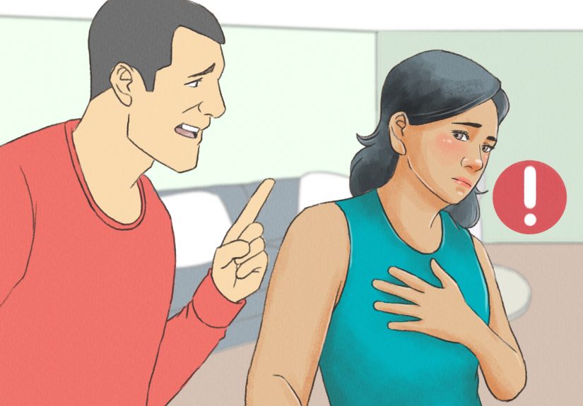
Caregivers also have their own experience of dysdiadochokinesia. They may see the person struggle with tasks that once looked automatic and wonder when to help and when to step back. Too much help can feel discouraging; too little can be unsafe. The best support often comes from learning the condition, reducing fall hazards, encouraging therapy, and recognizing that coordination problems are not laziness or lack of effort. They are neurologic.
Perhaps the most human part of the experience is this: people often mourn the loss of ease before they mourn the loss of ability. When ordinary movement no longer feels automatic, the day gets heavier. But many people also learn new strategies, adapt creatively, and rebuild confidence through therapy, pacing, and support. The movement may not be effortless anymore, but life can still be active, purposeful, and very much worth showing up for.
Final Thoughts
Dysdiadochokinesia is a neurologic sign that points to trouble with rapid alternating movement, most often because of cerebellar dysfunction or disruption of connected pathways. It can show up in stroke, multiple sclerosis, degenerative ataxias, toxic exposure, vitamin deficiency, trauma, infection, tumors, and other neurologic conditions. Diagnosis starts with the neurologic exam, but the real clinical task is identifying the underlying cause.
Treatment is usually cause-specific plus rehab-focused. Physical therapy, occupational therapy, speech therapy, adaptive tools, and fall-prevention strategies can all play major roles. Exercises may help improve control and function, but they work best when individualized and supervised. And if symptoms appear suddenly, urgent medical evaluation is the move. Not tomorrow. Not after one more cup of coffee. Now.