
Table of Contents >> Show >> Hide
- What is the cerebrum?
- Cerebrum diagram: a simple way to picture it
- Main parts of the cerebrum
- Functions of the cerebrum
- The lobes of the cerebrum and what they do
- Left brain vs. right brain: what is actually true?
- Cerebrum vs. cerebellum vs. brainstem
- What can affect the cerebrum?
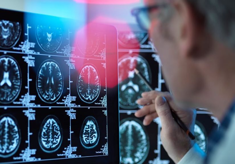
- How doctors evaluate cerebrum problems
- Everyday experiences related to the cerebrum
- Conclusion
- SEO Tags
The cerebrum is the brain’s big boss, idea factory, memory vault, and motion planner all rolled into one remarkably wrinkly package. If you’ve ever solved a math problem, recognized your best friend’s face, remembered where you left your keys, or decided not to text your ex back at 1 a.m., you can thank your cerebrum. It is the largest part of the brain, and it handles many of the high-level jobs that make you you.
In plain English, the cerebrum is the part of the brain that helps you think, feel, sense, move, communicate, learn, and adapt. It works with other brain structures, of course, but it does much of the heavy lifting when it comes to conscious awareness and complex behavior. That is why the cerebrum shows up in anatomy classes, neurology visits, brain scans, and just about every conversation about how the mind works.
This guide explains what the cerebrum is, what a simple cerebrum diagram shows, what each lobe does, how the two hemispheres differ, and what can happen when the cerebrum is injured or affected by disease. We will keep the science accurate, the wording human, and the jargon on a short leash.
What is the cerebrum?
The cerebrum is the largest part of the human brain. It sits at the top and front of the brain and is divided into two halves called the left hemisphere and right hemisphere. These hemispheres are connected by a thick bundle of nerve fibers called the corpus callosum, which lets the two sides share information quickly.
The outer layer of the cerebrum is called the cerebral cortex. This is the famous gray matter you see in brain illustrations. It is folded into ridges and grooves, which increases surface area without requiring your skull to be the size of a beach ball. Beneath the cortex lies white matter, which contains nerve fibers that connect different brain regions so messages can travel efficiently.
If you want the simplest possible definition, here it is: the cerebrum is the part of the brain responsible for most conscious thought and many higher functions, including voluntary movement, language, memory, reasoning, and sensory processing.
Cerebrum diagram: a simple way to picture it
A full medical illustration of the cerebrum can look a little like a walnut that enrolled in graduate school. To make things easier, here is a simplified text diagram showing the main lobes of the cerebrum. It is not to scale, but it gives you a useful mental map.
If you look at the cerebrum from above, you would see the left and right hemispheres separated by a deep groove. If you look from the side, the four major lobes are easier to identify: frontal at the front, parietal at the upper middle, temporal along the side, and occipital at the back. Some anatomy references also include the insula, a region tucked deeper inside the brain.
Main parts of the cerebrum
1. Left and right hemispheres
The cerebrum has two hemispheres, and each one helps control functions on the opposite side of the body. In other words, the left hemisphere generally helps control the right side of the body, while the right hemisphere generally helps control the left side. This crossing pattern is one reason a stroke on one side of the brain can affect movement or sensation on the opposite side.
2. Cerebral cortex
The cerebral cortex is the thin outer layer of gray matter. It is where many advanced tasks happen, including planning, attention, language, reasoning, and interpreting sensory information. It is also where much of your voluntary motor control begins.
3. White matter
White matter lies beneath the cortex and acts like the brain’s communication network. It links one brain area to another so signals can move fast and efficiently. If the cortex is the office staff, white matter is the hallway, Wi-Fi, and email system keeping everything connected.
4. Lobes
Each hemisphere is divided into lobes, and each lobe is associated with certain jobs. The key word here is associated, not exclusively responsible for. The brain works in networks, so most real-life tasks involve several regions at once.
Functions of the cerebrum
The cerebrum handles an impressive list of duties. Some are obvious, like helping you speak or move your hand. Others are more subtle, like helping you interpret a facial expression, stay focused during a meeting, or remember why you walked into the kitchen. Here are the major cerebrum functions:
- Voluntary movement: starting and guiding intentional body movements
- Sensory processing: interpreting touch, sight, sound, taste, and smell
- Language: producing speech, understanding words, reading, and writing
- Memory: storing and retrieving information
- Learning: building new skills and adapting based on experience
- Reasoning and problem-solving: comparing options, making decisions, and planning ahead
- Attention and awareness: focusing on what matters and filtering what does not
- Emotion and behavior: shaping responses, personality traits, and self-control
- Spatial awareness: understanding where your body is in relation to the world around you
That broad job description explains why the cerebrum is involved in so many neurological symptoms. Trouble with speech, weakness, confusion, personality change, vision loss, memory issues, seizures, or problems with attention can all point to cerebrum-related dysfunction, depending on which region is affected.
The lobes of the cerebrum and what they do
Frontal lobe
The frontal lobe sits at the front of the cerebrum and is often associated with executive function. That includes planning, judgment, decision-making, impulse control, and problem-solving. It also contains important motor areas that help initiate voluntary movement. In many people, parts of the frontal lobe on the left side also play a major role in speech production.
When the frontal lobe is affected, symptoms may include weakness, changes in behavior, reduced attention, poor judgment, or difficulty speaking. This is the lobe that helps you think before you act, which means it deserves a thank-you card more often than it gets.
Parietal lobe
The parietal lobe is located near the upper middle part of the cerebrum. It helps process sensory input, especially touch, pain, pressure, and body position. It also supports spatial awareness, which is how you know where your limbs are and how to move through space without constantly crashing into furniture.
Damage to the parietal lobe can lead to trouble recognizing objects, problems understanding spatial relationships, sensory changes, or difficulty paying attention to one side of the body or environment.
Temporal lobe
The temporal lobe sits along the side of the cerebrum near the temples. It is closely linked to hearing, language comprehension, memory, and emotion. Important structures involved in memory formation are associated with this region, which is why temporal lobe problems can affect how new memories are formed and stored.
Temporal lobe dysfunction may cause language comprehension problems, memory trouble, auditory processing issues, or seizures. In daily life, this can feel like hearing words but not quite decoding what they mean, which is frustrating and more common in neurological disorders than many people realize.
Occipital lobe
The occipital lobe is at the back of the cerebrum and is heavily involved in vision. It processes visual information such as shape, color, motion, and spatial detail. Your eyes collect light, but your occipital lobe helps turn that information into something meaningful.
If the occipital lobe is affected, symptoms may include vision changes, visual field loss, trouble recognizing what is being seen, or difficulty processing visual patterns.
Insula
The insula is a deeper region hidden within the folds of the cerebrum. It is often discussed less in beginner anatomy, but it matters. It appears to contribute to emotion, internal body awareness, taste, pain processing, and certain aspects of social and cognitive function. Think of it as one of the cerebrum’s quieter workers doing important background tasks without demanding the spotlight.
Left brain vs. right brain: what is actually true?
Popular culture loves to say the left brain is logical and the right brain is creative, as if your hemispheres are feuding roommates. Real neuroscience is more interesting and less dramatic. Some functions do tend to be more dominant on one side. For example, language is often more left-sided, especially in right-handed people, and some aspects of spatial processing are often more right-sided.
But most meaningful human tasks involve both hemispheres working together. Writing a song, solving a budget problem, driving a car, recognizing sarcasm, reading a map, and telling a joke are not single-lobe magic tricks. They rely on distributed brain networks communicating across the cerebrum.
So yes, lateralization is real. But no, you are not “100% right-brained” just because you own colored pens and have strong feelings about fonts.
Cerebrum vs. cerebellum vs. brainstem
These three structures are often mentioned together, and it helps to separate them clearly:
- Cerebrum: handles higher thinking, sensory interpretation, voluntary movement, language, memory, and emotion
- Cerebellum: helps coordinate movement, posture, and balance
- Brainstem: controls vital automatic functions such as breathing, heart rate, and wakefulness
All three are essential, but the cerebrum is the structure most people mean when they talk about thought, personality, and conscious processing.
What can affect the cerebrum?
Because the cerebrum is involved in so many functions, many conditions can affect it. Some are sudden, while others develop gradually.
Stroke
A stroke can interrupt blood flow to part of the cerebrum or cause bleeding in the brain. Depending on the affected region, symptoms may include facial drooping, arm weakness, speech problems, confusion, vision loss, numbness, or trouble walking. FAST is still a smart way to remember urgent stroke signs: Face drooping, Arm weakness, Speech difficulty, Time to call 911.
Traumatic brain injury
A blow to the head can injure the cerebrum and lead to headaches, confusion, memory issues, mood changes, difficulty concentrating, or loss of consciousness. Recovery varies widely based on the severity and location of the injury.
Seizure disorders and epilepsy
Abnormal electrical activity in the cerebrum can cause seizures. Symptoms may range from brief staring spells to involuntary movements, altered awareness, or unusual sensory experiences.
Brain tumors
Tumors in the cerebrum can cause headaches, seizures, weakness, speech changes, personality shifts, or visual symptoms. The exact signs depend heavily on location.
Dementia and neurodegenerative disease
Conditions that damage brain tissue over time may affect memory, reasoning, behavior, language, and daily function. Because the cerebrum supports so many higher-order abilities, it is often a major focus in disorders involving cognitive decline.
Infections and inflammation
Encephalitis and other inflammatory conditions can affect the cerebrum and lead to confusion, seizures, fever, weakness, or altered mental status. These situations need prompt medical care.
How doctors evaluate cerebrum problems
When a healthcare professional suspects a cerebrum-related issue, they usually begin with a detailed history and neurological exam. They may assess speech, strength, reflexes, sensation, vision, coordination, attention, and memory. Depending on the symptoms, testing may include:
- CT scan: often used quickly in emergencies, especially for head injury or possible stroke
- MRI: provides detailed images of brain tissue
- EEG: helps evaluate seizure activity
- Cognitive testing: measures memory, language, attention, and executive function
- Blood tests or lumbar puncture: may help if infection, inflammation, or metabolic problems are suspected
The key point is that symptoms help point to where the problem may be, while imaging and testing help explain why it is happening.
Everyday experiences related to the cerebrum
The cerebrum can sound abstract until you notice how often it runs the show in ordinary life. Imagine waking up and reaching for your phone before your eyes are fully open. Your occipital lobe helps process what you see on the screen, your temporal lobe helps you understand a voice note, your frontal lobe decides whether it is wise to answer messages before coffee, and your parietal lobe helps you coordinate your hand so you do not drop the phone on your face. That is not just morning chaos. That is brain anatomy in action.
Think about driving through a busy intersection. Your cerebrum is constantly collecting information, filtering distractions, remembering directions, interpreting traffic lights, monitoring speed, and preparing your muscles to move at the right moment. The frontal lobe helps with decision-making, the parietal lobe helps with spatial awareness, the occipital lobe handles visual input, and the temporal lobe helps you process sounds like a horn or siren. It is teamwork at a microscopic level, and it happens so fast that most people never notice it.
The cerebrum also shapes intensely personal experiences. If you have ever walked into a room and forgotten why, that is a reminder that attention and memory are not identical. If you have ever known the right word but could not get it out fast enough, you have had a tiny glimpse into how language networks can feel under pressure. If you have ever heard a song from high school and instantly felt a rush of memory, your temporal lobe would like some recognition for excellent archival work.
More serious cerebrum-related experiences can be unsettling. A person with a stroke affecting the left hemisphere might suddenly know what they want to say but find that the words come out tangled or not at all. Someone with a right parietal problem may bump into objects on one side and not understand why. A person with a frontal lobe injury may physically recover well but notice that planning, concentration, or emotional control feels different afterward. These are not character flaws. They are examples of how specific brain regions support specific parts of daily life.
Even recovery experiences often highlight the cerebrum’s remarkable adaptability. With therapy and practice, many people improve language skills after brain injury, strengthen attention after concussion, or relearn tasks after stroke. The brain is not infinitely flexible, but it does have an impressive ability to reorganize and build new pathways. Progress may be slow, messy, and deeply unglamorous, yet it is one of the most hopeful parts of neuroscience.
So when people ask what the cerebrum does, the best answer may be this: it helps turn sensation into meaning, intention into action, and experience into memory. It is not just a structure in an anatomy chart. It is the reason life feels like life.
Conclusion
The cerebrum is the largest part of the brain and one of the most important structures in the human nervous system. It includes two hemispheres, multiple lobes, a folded cerebral cortex, and an intricate communication network that supports movement, language, sensation, memory, emotion, and higher thinking. A simple cerebrum diagram can help you see where the frontal, parietal, temporal, and occipital lobes sit, but the real magic is in how these regions work together.
Understanding the cerebrum matters because it helps explain everything from reading and decision-making to stroke symptoms and brain injury recovery. In short, the cerebrum is not merely the biggest part of the brain. It is the part that gives daily life its meaning, flexibility, creativity, and complexity. Not bad for something that looks a little like a very ambitious walnut.